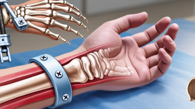

Fractures differ in location, shape, stability, and damage to nearby tissues. A clean break in the forearm may need a different approach from a shattered femur or an open ankle fracture. Fracture fixation methods are techniques used to keep broken bone ends aligned while healing takes place. The choice can affect movement, comfort, and the need for further treatment.
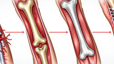
Some methods work from outside the body. External fixation uses pins placed into bone and connected to a frame, which can help stabilize serious injuries while wounds heal. Internal fixation places devices beneath the skin. Plates and screws can hold fragments in position, while an intramedullary nail sits inside the hollow center of a long bone. Wires or pins may support smaller bones or selected fracture patterns. Each option has trade-offs. No single method fits every fracture.
This guide introduces common fixation approaches and explains how clinicians consider them. Decisions often depend on the fracture’s pattern, bone quality, soft-tissue condition, and the patient’s overall health. Imaging and examination help inform that judgment. Treatment plans can change as an injury heals, and recovery timelines vary. Details matter. Even a technically successful repair may require careful rehabilitation and follow-up. This overview is educational, not a substitute for advice from an orthopedic professional who can assess the specific injury.
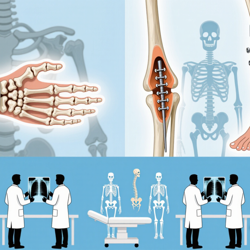
Fracture fixation holds broken bone ends in a useful position while healing begins. A cast or splint limits movement from outside the body. Internal fixation uses plates and screws, a rod inside the bone, or wires to steady fragments. External fixation uses pins connected to a frame outside the skin. The choice depends on the fracture’s location, pattern, skin condition, and the patient’s health. Small details matter: a screw placed near a joint can affect movement, while poor alignment may change how weight travels through the limb.
Stability does not mean keeping every fracture completely motionless. Surgeons aim for enough support to protect alignment while the bone forms new tissue. Too much movement can disturb healing; excessive rigidity may also be undesirable in some fracture patterns. It is a balance, not a simple rule. The GBD 2019 Fracture Collaborators estimated 178 million new fractures worldwide in 2019, in a report published in The Lancet Healthy Longevity. That figure shows the scale of fracture care, but it does not prove one fixation method works best. Hardware is not magic. Follow-up images and clinical checks help clinicians judge whether alignment and healing are progressing, and plans may need adjustment.
| Fixation method | How it stabilizes the fracture | Common uses | Key considerations |
|---|---|---|---|
| Plate-and-screw fixation | A metal plate is secured to the bone with screws to hold fragments in alignment. Depending on the fracture and construct, it can provide compression or bridge a fracture zone. | Many fractures near joints, fractures with several fragments, and fractures that need direct alignment, including some fractures of the forearm, wrist, and lower leg. | Often allows stable alignment, but usually requires an operation and exposure of the fracture. The appropriate plate position and fixation depend on the bone and fracture pattern. |
| Intramedullary nail | A rod is inserted into the central canal of a long bone and typically locked with screws at one or both ends to resist rotation and shortening. | Many shaft fractures of long bones, especially the femur and tibia; selected humeral shaft fractures may also be treated this way. | It can stabilize the bone while preserving much of the surrounding soft tissue. The entry point, fracture pattern, and bone anatomy influence whether it is suitable. |
| External fixation | Pins or wires are placed into bone and connected to a frame outside the body. The frame holds the bone in position without placing the main stabilizing structure under the skin. | Severe open fractures, major soft-tissue injury, some pelvic fractures, and situations where temporary stabilization is needed before further treatment. | Can provide access to injured soft tissues, but pin sites require care and infection can occur. It may be temporary or, in selected cases, definitive treatment. |
| Screw fixation | One or more screws hold bone fragments together. A screw may compress fragments or secure a fragment to a larger bone segment, depending on its design and placement. | Selected fractures with a simple pattern, such as some fractures around joints or small bone fragments that can be securely captured. | Works best when the fracture pattern and bone quality allow secure placement. Screws may also be used together with plates or other fixation devices. |
| Kirschner wires or pins | Thin metal wires or pins are passed through bone to hold smaller fragments in position, often as part of a percutaneous or minimally invasive procedure. | Some fractures of the fingers, hand, wrist, and other small bones, particularly when fragments are relatively small. | May be used temporarily or as definitive fixation. Stability varies by fracture and wire configuration; exposed wire ends need appropriate pin-site care. |
| Tension-band fixation | A wire or cable construct converts forces that tend to pull a fragment apart into compression across the fracture during movement. | Selected fractures where muscle pull tends to separate a fragment, such as certain kneecap or elbow-tip fractures. | Suitable only for specific fracture patterns. The construct must be positioned appropriately, and irritation from prominent hardware can occur. |
| Cast or splint immobilization | External support limits movement and helps maintain alignment while the bone heals; it does not use implanted hardware. | Stable fractures, many fractures in children, or fractures that can be held in acceptable alignment without surgery. | Alignment and healing need follow-up. Swelling, skin pressure, and stiffness are considerations, and some fractures may shift despite immobilization. |
How fixation supports healing: Fixation limits unwanted movement and helps maintain bone alignment. The best method depends on factors such as fracture location and pattern, soft-tissue condition, bone quality, and the patient’s overall health. A qualified clinician determines the treatment plan for an individual fracture.